
May 23, 2013
Imagine holding a leaf more than a foot from your face and being able to examine its fine details on the scale of micrometres or even nanometres.
This kind of high-resolution far field imaging is one step closer to realization thanks to the work of Professor George Eleftheriades and PhD candidate Alex Wong of The Edward S. Rogers Sr. Department of Electrical & Computer Engineering.
Have you ever wondered why you can’t look through a magnifying glass and see individual molecules? It’s partly because of a property of light called diffraction that limits its resolution. For visible light, the kind that lets you see through a normal magnifying glass, the diffraction limit depends on two factors: the wavelength of the wave, and the distance from the object you want to see.
Historically, there were only two ways around the diffraction limit to see high resolution images: use waves with a shorter wavelength, such as x-rays or electron beams, or put a probe extremely close to the object you’re trying to see—within a few nanometres for visible light. With a microscope, you’re ultimately limited by the wavelength of visible light—in the same way that you can’t trace a line thinner than the tip of the pen you’re using, Eleftheriades explains.
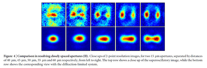
Wong and Eleftheriades have created a super-microscope that allows optical imaging beyond the diffraction limit, without using short-wavelength waves or getting really close and without the need for fine scanning. Their work was recently published in the Nature journal Scientific Reports.
How? About four years ago, Eleftheriades and Wong saw a connection between their work and an old antenna theory that had largely been abandoned. By modifying the theory to their problem, they were able to create a superoscillation-a tailored rapid variation in a wave’s shape—allowing them to locally ‘shrink’ the wavelength beyond the diffraction limit.
They first demonstrated their ability to deterministically design and launch a superoscillation in radio waves, earning them the IEEE’s 2012 R.W.P. King Award for best paper in the journal IEEE Transactions on Antennas and Propagation.
The advantage with their system is its simplicity of use, low cost and ability to image in real time. Current methods of “super-resolution” microscopy in biology and medicine often require pre-treating the specimen with fluorescent tags or dyes, or using oil immersion techniques to increase magnification. Sub-diffraction imaging requires none of that mess, fuss or complex set up.
Apart from life sciences the super-microscope also has potential applications in manufacturing—for example, allowing manufacturers to see miniscule defects or roughness on surfaces in real time, without having to get nanoscale-close. It could also be used by the microelectronics industry to perform quality control checks quickly and without having to finely scan.
Wong and Eleftheriades hope to apply their system as a component in a conventional optical microscope. “I want to make it more general,” says Eleftheriades. “It’s just a matter of designing it better with stronger lasers.”
For Wong, arriving at a functioning prototype after five years has been the cherry on top of his PhD. “I was fortunate to be involved in a project and get to this last step,” says Wong. “There’s a strong feeling of accomplishment—it’s exciting and satisfying, but there’s always room for improvement.”
Eleftheriades also sees their achievement as one component in a larger effort. “We do a step, someone else does another step, but always science wins,” he says.
More information:
Marit Mitchell
The Edward S. Rogers Sr. Department of Electrical & Computer Engineering
416-978-7997; marit.mitchell@utoronto.ca